Amsler Grid
A chart made of lines that make up a grid pattern used to assess central vision and therefore how well the macula functions. Please click this
link to obtain a grid from the internet together with instruction on its use.
Anaesthesia
Local anaesthesia is an injection of a drug around the eye that numbs the eye and enables surgery to be performed without discomfort whilst the patient is awake. General anaesthesia is when the patient is given drugs that make them fall asleep for the operation.
Astigmatism
A condition where the front surface of the cornea (clear window at the front of the eye) is more curved in one direct that the other (like a rugby ball). It is corrected by glasses or contact lenses but can also be corrected at the time of cataract surgery by use of a toric lens.
Biometry
A measurement of the curvature of the front of the eye and the length of the eye that allows calculation of the strength of lens required at the time of cataract surgery.
Blepharitis
The most common disease of the eyelids. It is a sterile low grade inflammation of the lid margins (where the lashes are situated) mainly treated with simple lid hygiene.
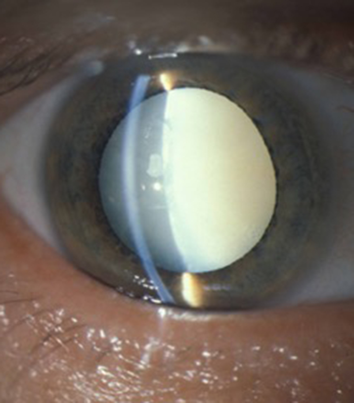
Cataract
A cloudy lens inside the eye which usually occurs as you get older and requires a surgical procedure to correct. It causes gradual blurring of vision in one or both eyes and can be associated with glare symptoms.
Cornea
The clear window at the front of the eye and is the main structure that allows light rays to focus on the retina. This is where contact lenses are placed to correct problems with the focus of the eye.
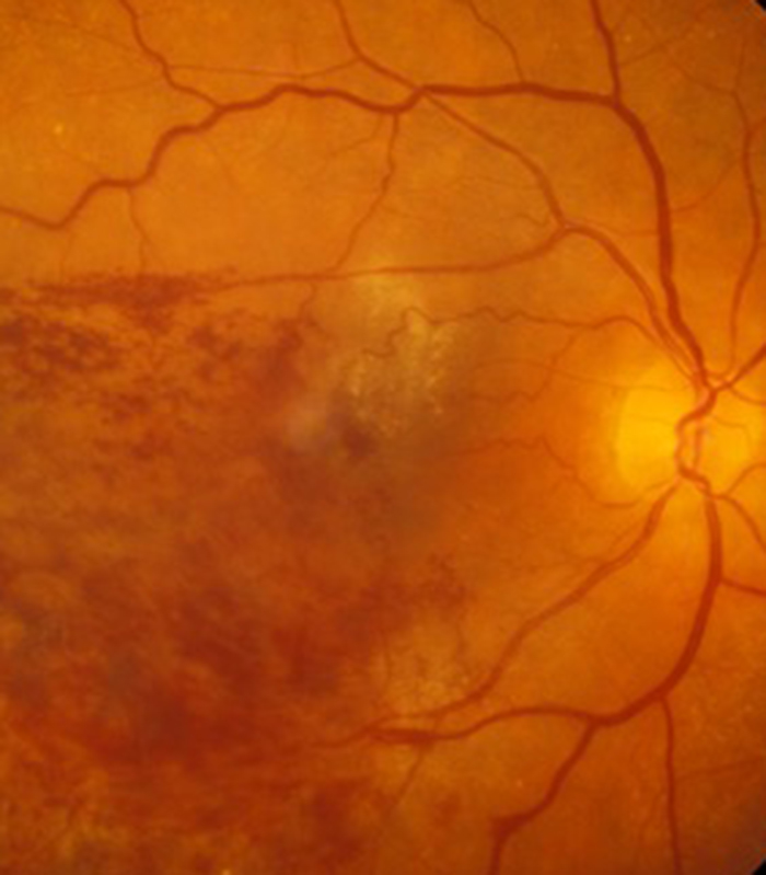
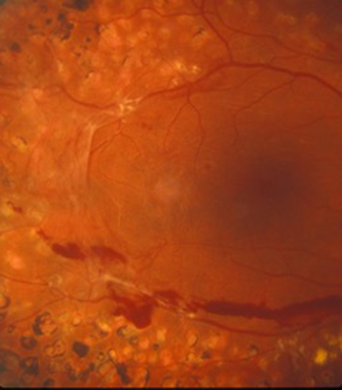
Diabetes
This condition occurs when the blood sugar is too high and can result in problems with circulation of the retina with the passage of time. The problem is with the small blood vessels (capillaries) which can either block (called ischaemia) or leak fluid (called oedema).
Fluorescein angiography
An accurate way of assessing the circulation in the retina. It may be used in macular degeneration, diabetic eye disease and retinal vein occlusion. Iodine dye is injected into a vein in the arm and then photographs are taken of the back of the eye to see how the dye circulates as it passes through the retina.
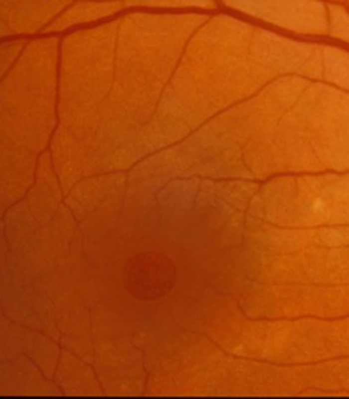
Fovea
The very centre of the macula. This area gives us the best quality vision and colour vision. This is the retina that is used when you look directly act something.
Glaucoma
When the pressure in the eye is too high for the eye and can damage the optic nerve that transmits the messages from the eye to the brain.
Hypermetropia (long sight)
The eye is too short for the lenses at the front of the eye and cannot focus light on the retina. If this condition is mild and in younger patients the crystalline lens inside the eye (which can change shape to increase focus) can accommodate (increase its focus) to keep the image on the retina. As you get older this accommodation ability reduces and eventually image is not longer in focus and you need glasses to be worn for distance and an even stronger lens is needed for near.
Intraocular lens
The small plastic lens that is inserted into the eye at the time of cataract surgery to replace the focus that is lost when the cataract is removed.
Intraocular pressure
A clear fluid called aqueous circulates inside the eye and maintains the eye at a pressure above atmospheric pressure called the intraocular pressure.
Laser
A particular type of light that can be used both for diagnosis at low powers (to measure the eye) and treatment at high powers (to treat the posterior capsule and retina)
Lens (of eye)
The lens inside the eye (crystalline lens) is the variable fine focus of the eye (compared to the fixed coarse focus of the cornea) and until late middle age can change its shape to enable us to both focus into the distance (at the horizon) when the lens is in its relaxed shape and near (for reading) when accommodation occurs and the lens changes shape (thickens) to increase the focusing power for near to enable us to read a book. This power of accommodation is at its maximum at the age of 18 then starts to decline. Fortunately there is plenty in reserve so we do not need reading glasses until we are usually in our 40's or 50's.
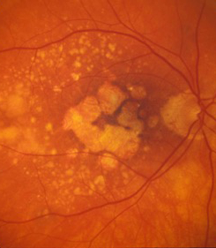
Macula
The central area of the retina used when we look directly at something. At its very centre is the fovea.
Monovision
A condition created at the time of cataract surgery when both eyes are having surgery with one eye focused for distance and one eye focused for near
Multifocal lenses
A lens inserted into the eye at the time of cataract surgery that has multiple focus with some light being focused for near, intermediate distance and the far distance.
Myopia (short sight)
The eye is too big for the lenses in the eye and they focus the light in front of the retina. The vision is blurred in the distance but closer objects may be in focus. Glasses are needed for the distance.
OCT
Optical Coherence Tomography is a laser scanner that can take detailed images of the various laser of the retina.
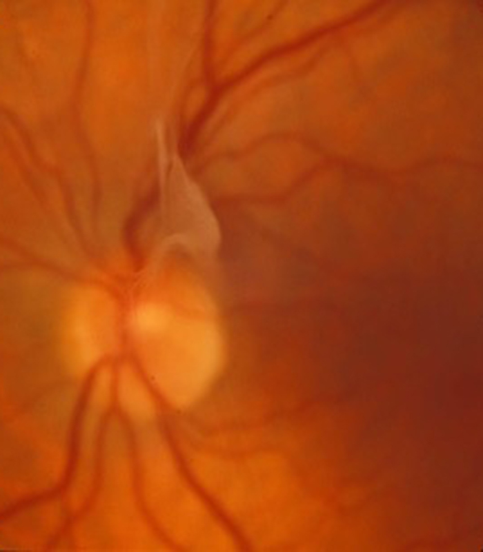
Optic nerve
The nerve that collects all the electrical information from the retina and sends it to the brain to give us sight.
Phacoemulsification
This is the modern method of removing a cataract. The phacoemulsifier is a thin tube that vibrates a small amount but at great speed. This breaks the cataract into many small pieces that get sucked up the tube and out of the eye.
Phacovitrectomy
This is a “combined” operation when the lens of the eye is removed at the same time as performing a vitrectomy. This avoids the patient having to return for further surgery for cataract months or years after the vitrectomy.
Posterior capsule
A thin clear membrane left behind at the time of cataract surgery to support the new lens that is placed inside the eye.
Povidone-iodine
A type of iodine solution that the most effective thing to quickly destroy bacteria and is used to clean the skin around the eye before surgery.
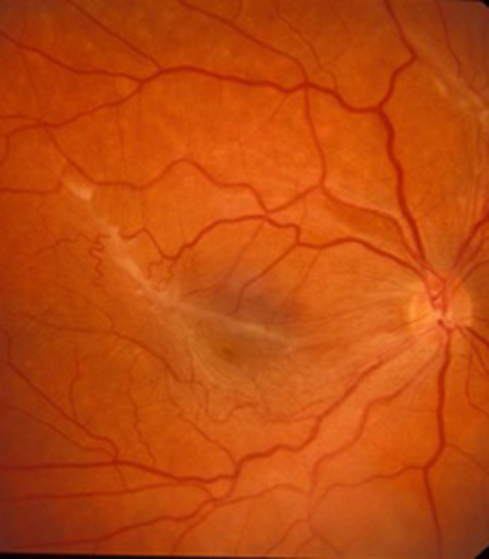
Retina
Photographic film at the back of the eye. Cells called rods and cones in the retina convert light into electricity which is then sent to the brain by the optic nerve to give us sight.
Toric lenses
A type of lens inserted at the time of cataract surgery to correct astigmatism.
Vitreous
The clear jelly that occupies the main eye cavity.
Vitrectomy
Removal of the vitreous from the eye. This may be required for surgery to allow access to the retina which lines the back wall of the eye.